Modern Dermoscopy.
Recent literature, society guidelines, and conference content, organized by clinical domain. Continuously updated.
About this resource
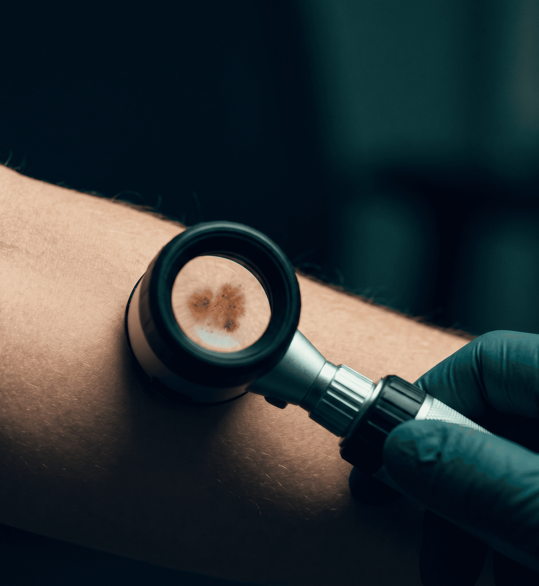
Dermoscopy Updates is an editorially curated reference covering modern dermoscopy practice. Each topic merges current peer-reviewed literature, major conference content, and practice changes into a tight, citation-backed brief. High yield clinical points first, recent updates second, full source attribution always.
Sections
Foundations
The dermoscopic alphabet that lets you read every other lesion: light, structures, vessel morphologies, and color cues.
Diagnostic Methods & Management
Pattern analysis, the 7-point and 3-point checklists, comparative approach, and the Lallas management rules.
Melanocytic Lesions
Nevus pattern catalog, Spitz/Reed lesions, blue and combined nevi, congenital melanocytic nevi.
Melanoma
From pattern analysis through superficial spreading, lentigo maligna, acral, and rare melanoma subtypes (nodular, amelanotic, spitzoid).
Actinic Keratosis & Squamous Cell Carcinoma
The continuum from photodamaged macule through SCCIS / Bowen, well-differentiated SCC, high-risk SCC, mucosal in-situ disease (PeIN), and clinical mimickers.
Basal Cell Carcinoma
Five subtypes with distinct dermoscopic signatures: nodular, superficial, pigmented, morpheaform, plus a unifying overview.
Other Cutaneous Malignancies
Merkel cell carcinoma, atypical fibroxanthoma / pleomorphic dermal sarcoma, dermatofibrosarcoma protuberans, Kaposi sarcoma, cutaneous T-cell lymphoma.
Benign Tumors
Seborrheic keratosis, dermatofibroma and fibrohistiocytic lesions, the full vascular lesion spectrum.
Inflammoscopy
Vessels, scale, color: psoriasis, eczema, lichen planus, granulomatous and autoimmune disease, infections, and early mycosis fungoides.
Special Sites
Nail unit (onychoscopy), volar skin (palms and soles), vulvar mucosal dermoscopy.
Hair & Scalp
Trichoscopy of androgenetic alopecia and alopecia areata, plus scalp pathology beyond trichoscopy (CCCA, scalp DLE, scarring alopecias).
Imaging Modalities
Reflectance confocal microscopy: cellular-level imaging on equivocal lesions.
Practice & Self-Test
Case-based review and pattern-recognition drills.
Foundations
The dermoscopic alphabet that lets you read every other lesion: light, structures, vessel morphologies, and color cues.
Diagnostic Methods & Management
Pattern analysis, the 7-point and 3-point checklists, comparative approach, and the Lallas management rules.
Melanocytic Lesions
Nevus pattern catalog, Spitz/Reed lesions, blue and combined nevi, congenital melanocytic nevi.
Melanoma
From pattern analysis through superficial spreading, lentigo maligna, acral, and rare melanoma subtypes (nodular, amelanotic, spitzoid).
Actinic Keratosis & Squamous Cell Carcinoma
The continuum from photodamaged macule through SCCIS / Bowen, well-differentiated SCC, high-risk SCC, mucosal in-situ disease (PeIN), and clinical mimickers.
Basal Cell Carcinoma
Five subtypes with distinct dermoscopic signatures: nodular, superficial, pigmented, morpheaform, plus a unifying overview.
Other Cutaneous Malignancies
Merkel cell carcinoma, atypical fibroxanthoma / pleomorphic dermal sarcoma, dermatofibrosarcoma protuberans, Kaposi sarcoma, cutaneous T-cell lymphoma.
Benign Tumors
Seborrheic keratosis, dermatofibroma and fibrohistiocytic lesions, the full vascular lesion spectrum.
Inflammoscopy
Vessels, scale, color: psoriasis, eczema, lichen planus, granulomatous and autoimmune disease, infections, and early mycosis fungoides.
Special Sites
Nail unit (onychoscopy), volar skin (palms and soles), vulvar mucosal dermoscopy.
Hair & Scalp
Trichoscopy of androgenetic alopecia and alopecia areata, plus scalp pathology beyond trichoscopy (CCCA, scalp DLE, scarring alopecias).
Imaging Modalities
Reflectance confocal microscopy: cellular-level imaging on equivocal lesions.
Practice & Self-Test
Case-based review and pattern-recognition drills.
Bottom line
Dermoscopy moves fast. New diagnostic criteria, AI tools, biopsy alternatives, and treatment guidelines land every year. This resource is built so a working clinician can stay current in minutes per topic, not hours.
Pick a section. Everything you need is on one page per topic.